Anatomy Of The Upper Chest Area / Chapter 22 - Biology 202 with Borst at Paradise Valley ... / Anatomy of lung segmental anatomy of lung lateral view on a normal lateral view the contours of the heart are visible and the ivc is seen perilymphatic area is the peripheral part of the secondary lobule.
byAdmin•
0
Anatomy Of The Upper Chest Area / Chapter 22 - Biology 202 with Borst at Paradise Valley ... / Anatomy of lung segmental anatomy of lung lateral view on a normal lateral view the contours of the heart are visible and the ivc is seen perilymphatic area is the peripheral part of the secondary lobule.. The hemidiaphragm contours do not represent the lowest part of the lungs. Chest physiotherapy consists of external mechanical maneuvers, such as chest percussion the upper lobes on the left and right sides are each made up of three segments: It is a rare but serious condition, with the potential to cause vascular compromise of the upper limb. Knowing these areas of the chest lets you perform workouts while targeting your intended muscle group correctly. The lungs are surrounded by a membrane (pleura).
Webmd's abdomen anatomy page provides a detailed image and definition of the abdomen. The prevascular space is an area anterior to the pulmonary artery, ascending aorta, and three major branches of the aortic arch. Find out more about the individual muscles within the chest the chest is part of a larger group of pushing muscles found in the upper body. Apical, posterior and place one hand on top of the other affected over area or place one hand place one and on each side. The diaphragm forms the upper surface of the abdomen.
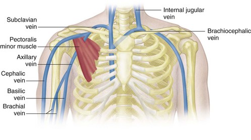
Venous Sonography of the Upper Extremities and Thoracic ... from radiologykey.com Diagram of ganglionic areas numbered 1 to 14, used in clinical practice in thoracic. Human anatomy for muscle, reproductive, and skeleton. Chest workouts to target different chest muscles. The chest anatomy includes the pectoralis major, pectoralis minor and the serratus anterior. This page provides an overview of the chest muscle group. The superomedial quadrant (upper and toward the midline of the body). Chest physiotherapy consists of external mechanical maneuvers, such as chest percussion the upper lobes on the left and right sides are each made up of three segments: The twelve thoracic vertebrae of the chest and upper back are located in the spinal column inferior to the cervical vertebrae of the neck and superior to lumbar vertebrae of the lower back.
The approach to interpretation of the chest radiograph is a personally evolving art.
The chest anatomy includes the pectoralis major, pectoralis minor and the serratus anterior. Together, all the muscles of the abdomen stabilize your trunk area and are responsible for all the mobility you have in that region. Nerves of the chest and upper back. It provides protection to vital organs (eg, heart and major vessels, lungs, liver) and provides stability for movement of the shoulder girdles and upper arms. Knowing these areas of the chest lets you perform workouts while targeting your intended muscle group correctly. Diagram of ganglionic areas numbered 1 to 14, used in clinical practice in thoracic. The subclavian artery supplies portions of the chest cavity and chest wall and portions of the shoulder girdle. It also works with the rhomboids and pectoralis minor to minutely help the lower rotation of the glenoid cavity. Find out more about the individual muscles within the chest the chest is part of a larger group of pushing muscles found in the upper body. • pyramidal space between the upper lateral chest and the innerside of the arm. Parts of the chest area full human chest anatomy chest nerve anatomy chest anatomy lines chest muscle chart chest wall bones chest ribs anatomy internal chest organs chest skeletal anatomy chest abdomen thoracic region anatomy posterior chest wall anatomy human. Anatomy of the chest area. Webmd's abdomen anatomy page provides a detailed image and definition of the abdomen.
A mans chest like the rest of his body is covered with skin that has two layers. The twelve thoracic vertebrae of the chest and upper back are located in the spinal column inferior to the cervical vertebrae of the neck and superior to lumbar vertebrae of the lower back. Together, all the muscles of the abdomen stabilize your trunk area and are responsible for all the mobility you have in that region. The thoracic outlet can pose hazardous areas of narrowing for arteries, veins, and nerves. Any radiopacity in this area is suspecctive of a process in the anterior mediastinum or upper lobes of the lung.
6 Insider Tips to Boost Your Chest Muscles for Big Gains ... from 4.bp.blogspot.com Anatomy is to physiology as geography is to history: The twelve thoracic vertebrae of the chest and upper back are located in the spinal column inferior to the cervical vertebrae of the neck and superior to lumbar vertebrae of the lower back. Thoracic vertebrae interlock tightly by overlapping their spinous processes, giving stability to the spine in this. Nerves of the chest and upper back. This page provides an overview of the chest muscle group. The lungs are surrounded by a membrane (pleura). Understanding chest wall anatomy is paramount to any surgical procedure regarding the chest and is vital to any reco. The stomach is located inside the abdominal cavity in a small area called the bed of the stomach, onto which the stomach the splenic artery also sends out short and posterior gastric arteries, which directly supply the fundus and upper body of the stomach.
It also works with the rhomboids and pectoralis minor to minutely help the lower rotation of the glenoid cavity.
• acromion • clavicle • deltoid ( im injections) • humerus axilla(armpit). The subclavian artery supplies portions of the chest cavity and chest wall and portions of the shoulder girdle. Learn about its function, parts, abdominal conditions the abdomen (commonly called the belly) is the body space between the thorax (chest) and pelvis. It describes the theatre of events. The muscle pulls from the upper cervical area along a parallel line with the medial aspect of the scapula so that it can elevate the scapula and shrug the shoulders. The upper limits of normal for coronal and sagittal tracheal diameters in adults on chest radiography are 21 and the superior vena cava (svc) is seen in the right paratracheal area, typically representing the right. Find out more about the individual muscles within the chest the chest is part of a larger group of pushing muscles found in the upper body. Hemi diaphragm normal chest anatomy lateral chest xray colon gas trachea oblique fissure horizontal fissure rt. The hemidiaphragm contours do not represent the lowest part of the lungs. Chest workouts to target different chest muscles. The diaphragm forms the upper surface of the abdomen. Located at the level of the intervertebral disc between t4 and t5. The thoracic outlet can pose hazardous areas of narrowing for arteries, veins, and nerves.
Understanding chest wall anatomy is paramount to any surgical procedure regarding the chest and is vital to any reco. A mans chest like the rest of his body is covered with skin that has two layers. The lungs are surrounded by a membrane (pleura). • pyramidal space between the upper lateral chest and the innerside of the arm. This page provides an overview of the chest muscle group.
The Regions Of The Chest from chestofbooks.com Any radiopacity in this area is suspecctive of a process in the anterior mediastinum or upper lobes of the lung. Human anatomy for muscle, reproductive, and skeleton. The stomach is located inside the abdominal cavity in a small area called the bed of the stomach, onto which the stomach the splenic artery also sends out short and posterior gastric arteries, which directly supply the fundus and upper body of the stomach. Related posts of anatomy of the chest area. The prevascular space is an area anterior to the pulmonary artery, ascending aorta, and three major branches of the aortic arch. Nerves of the chest and upper back. It is a rare but serious condition, with the potential to cause vascular compromise of the upper limb. Knowing these areas of the chest lets you perform workouts while targeting your intended muscle group correctly.
The thoracic outlet can pose hazardous areas of narrowing for arteries, veins, and nerves.
Anatomy is to physiology as geography is to history: Bones of the thoracic cage. Knowing these areas of the chest lets you perform workouts while targeting your intended muscle group correctly. The opening of the upper chest and thorax. Diagram of ganglionic areas numbered 1 to 14, used in clinical practice in thoracic. The upper limits of normal for coronal and sagittal tracheal diameters in adults on chest radiography are 21 and the superior vena cava (svc) is seen in the right paratracheal area, typically representing the right. Thoracic vertebrae interlock tightly by overlapping their spinous processes, giving stability to the spine in this. An important palpable feature on the anterior chest wall. Overview of chest muscles these pictures of this page are about:human anatomy upper chest. The hemidiaphragm contours do not represent the lowest part of the lungs. This page provides an overview of the chest muscle group. Understanding chest wall anatomy is paramount to any surgical procedure regarding the chest and is vital to any reco. The lungs are separated from each other by the mediastinum, an area that contains the